August 1, 2015
Mentor – Dr. Mukesh Taneja
Other Contributors – Amulya Kishore, Sravan L., Badrinath Singhal
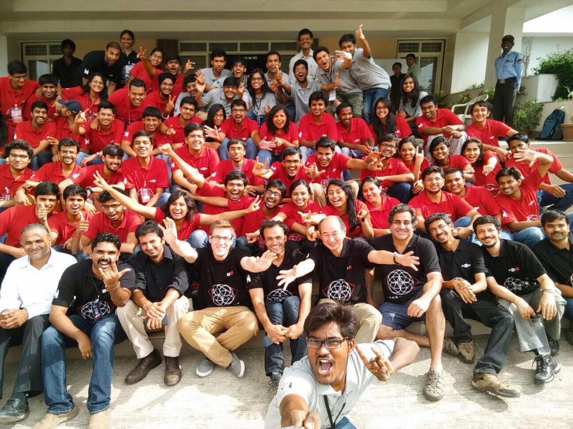
During the summer of 2015, I got an opportunity to be a part of the ReDx Engineering the Eye program jointly organized by the Massachusetts Institute of Technology Media Lab (Camera Culture group) and the L V Prasad Eye Institute in Hyderabad, India. I was one of the 63 participants selected for the event from all across the country (India). During this week long workshop, we (a team of four) created to low-cost system to image the meibomian glands (present on the inner side of our eye lid) and detect their deficiency which leads to a chronic condition – Dry Eye Syndrome. The following poster briefly describes our work.
My main responsibility was to create the imaging hardware suitable for the application. We experimented with a variety of image sensors ranging from a ultra high resolution night camera to a basic desktop webcam with the aim of balancing image quality with low cost (which was an important concern considering the scalability goals of the project). Owing to its high skin penetration capacity, Near Infra-Red (NIR) proved to be the appropriate illumination source. We hacked into a desktop webcam to make it sensitive to IR (and less sensitive to other wavelengths). Apart from this, the project provided me my first experience at CAD designing and 3-D printing.